
Articular cartilage injury can sometimes be confusing, because there are three different types of cartilage found in the body: articular or hyaline cartilage (covers joint surfaces), fibrocartilage (knee meniscus, vertebral disk), and elastic cartilage (outer ear). These different cartilages are distinguished by their structure, elasticity, and strength.
Articular cartilage is a complex, living tissue that lines the bony surface of joints. Its function is to provide a low friction surface enabling the joint to withstand weight bearing through the range of motion needed to perform activities of daily living as well as athletic endeavors. In other words, articular cartilage is a very thin shock absorber. It is organized into five distinct layers, with each layer having structural and biochemical differences.
What causes an articular cartilage injury?
Articular cartilage injuries can occur as a result of either traumatic injury or progressive degeneration over time (wear and tear). A direct blow or other trauma can injure the articular cartilage. Articular cartilage has no direct blood supply, thus it has little or no capacity to repair itself. If the injury penetrates the bone beneath the cartilage, the underlying bone provides some blood to the area, improving the chance of healing. Injury to the articular cartilage is often associated with swelling in the affected joint and pain that can wax and wane over time. In severe cases where the cartilage is sheared off of the surface, mechanical symptoms such as locking and catching can occur.
Mechanical degeneration (wear and tear) of articular cartilage occurs with the progressive loss of the normal cartilage structure and function. This initial loss begins with cartilage softening then progresses to fragmentation. As the loss of the articular cartilage lining continues, the underlying bone has no protection from the normal wear and tear of daily living and begins to break down, leading to osteoarthritis.
What are the symptoms of articular cartilage injury?
In many cases, a patient will experience knee swelling and vague pain. At this point continued activity may not be possible. If a loose body is present, words such as “locking” or “catching” might be used to describe the problem.
With wear and tear, the patient often experiences stiffness, decreased range of motion, joint pain, and/or swelling.
How is an articular cartilage defect (injury) diagnosed?
The physician examines the knee, looking for decreased range of motion, pain along the joint line, swelling, fluid on the knee, abnormal alignment of the bones making up the joint, and ligament or meniscal injury. Injuries to the articular cartilage are difficult to diagnose, and evaluation with MRI (magnetic resonance imaging) or arthroscopy (performing a surgery to look inside the joint with a small camera) may be necessary. Plain X-rays are not usually good in diagnosing articular cartilage problems but are usually taken to rule out other abnormalities.
How is an injury treated?
Injuries to the cartilage that do not extend to the bone will generally not heal on their own. Injuries that penetrate to the bone may heal, but the type of cartilage that is laid down is different than the injured cartilage – it is structurally unorganized and does not function as well as the original articular cartilage.
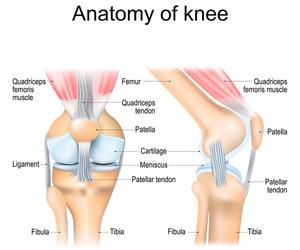
For smaller articular cartilage defects which are asymptomatic, surgery may not be required. Defects smaller than 2 cm have the best prognosis and treatment options. These include arthroscopic surgery using techniques to remove damaged cartilage and increase blood flow from the underlying bone (e.g., microfracture) or transplanting cartilage from other areas of the knee (osteochondral autograft transplantation). For larger defects, the options range from growing ones own cells and implanting them into the defect (autologous chondrocyte implantation) to using a donated pieced of cartilage and bone from a cadaver (osteochondral allograft transplantation). If you have a cartilage injury, consult your specialist for further information on the decision to have surgery and what type of procedure would be best for you.
Reviewed June 2026 by Elizabeth Dennis, MD
Contributing experts: Bert R. Mandelbaum, MD; Daniel Romanelli, MD; John Uribe, MD; Diane S. Watanabe, ATC
Sports Tips are brought to you by the American Orthopaedic Society for Sports Medicine. They provide general information only and are not a substitute for your own good judgment or consultation with a physician.
For more tip sheets and to find an expert near you, visit sportsmed.org/STOP
Copyright ©2025. American Orthopaedic Society for Sports Medicine. All rights reserved.